
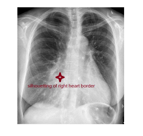
The frontal chest x-ray shows a zone of opacification silhouetting the right heart border.
The lateral chest x-ray shows abnormal chest wall with severe pectus excavatum.

Silhouetting of the right heart border can be due to 3 causes.
- Right middle lobe consolidation.
- Right middle lobe collapse.
- Pectus excavatum due to displacement of the heart by the depressed sternum.
Bottom line is frontal chest x-ray in a patient with pectus excavatum can simulate right middle lobe disease.
With thanks to Dr. Faheem Afzal for the images.
Reference: Grainger & Allison’s Diagnostic Radiology, 6th Edition.