
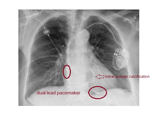
The chest x-ray shows dual chamber pacemaker with leads projected over the right atrium and the right ventricle. There is a curvilinear calcified area in the lower left para vertebral area of the heart indicating the presence of mitral annular calcification.
A dual chamber pacemaker has leads in the right atrium and the right ventricle. Right atrial lead is usually placed in the appendage which is located superiorly in the right atrium. This appears on the lateral chest x-ray with the right atrial lead curving upwards and anteriorly.
The right ventricular lead tip should be to the left of the spine on the frontal chest x-ray. On the lateral view, the lead should course anteriorly into the apex of right ventricle.
Mitral annular calcification (MAC) is seen in elderly patients. It is believed to be secondary to chronic degeneration of the fibrous ring of the mitral valve and may be seen in younger patients with abnormal calcium metabolism. It is seen as O or C shaped dense structure at the expected location of the mitral annulus.
Reference : Brant and Helms’ Fundamentals of Diagnostic Radiology, 5th Edition & https://pubs.rsna.org/doi/full/10.1148/rg.316115529