
The frontal wrist x-ray shows a scaphoid fracture. There is crowding of the carpal bones as opposed to what should be a normal configuration with 1-2 mm distance between the carpal bones.

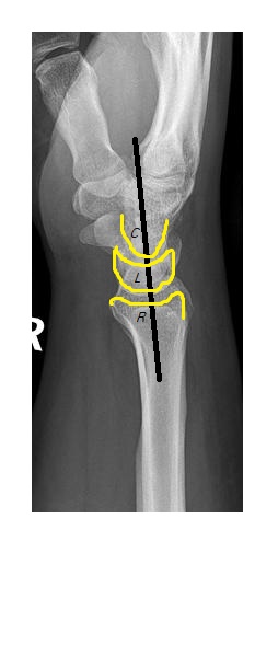
On the lateral view, the capitate along with the metacarpals are dislocated posteriorly with resultant perilunate dislocation.

The patient underwent operative management.
Perilunate dislocation:
- Common mechanism is fall on outstretched hand.
- Ligament disruption between the capitate & lunate allows the capitate to dislocate from the cup shaped distal lunate surface.
- On Ap view, the lunate, instead of showing a normal rhomboid shape with parallel upper and lower borders, appears triangular or pie shaped.
- On the lateral view, there is disruption of the normal cup & saucer anatomy between distal radius, lunate & capitate.
- Perilunate dislocation is commonly associated with a transscaphoid fracture.
Reference : Fundamentals of Diagnostic Radiology by Brant & Helms.