
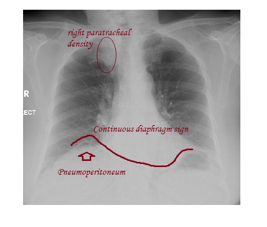
The technically poor AP erect chest x-ray shows air under the right hemidiaphragm consistent with a pneumoperitoneum.
The patient had a CT scan of the abdomen which showed a sigmoid diverticular perforation and was managed surgically.
The right paratracheal density seen as an incidental finding was related to venous confluence.
Continuous diaphragm sign is seen when there is large amount of intraperitoneal free air extending across the undersurface of the diaphragm.
Reference: Essentials of Pediatric Radiology: A Multimodality approach.