A 17 year old presents with right knee pain and swelling following a motor vehicle accident. What does the knee x-ray show?
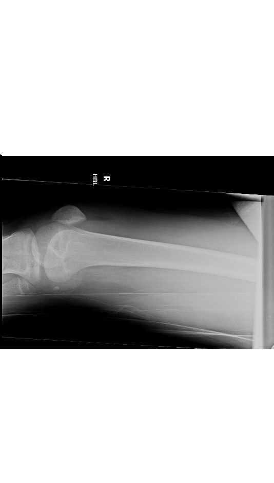
The knee xray shows lipohaemarthrosis which is essentially a fat blood interface. It indicates an intra articular fracture around the knee joint. Even if a fracture is not identified immediately ,one should assume that an undisplaced fracture is present ( similar to elbow fat pad or sail sign)
Fat released from the marrow layers on top of blood ( effusion ) in the suprapatellar bursa. Only seen on lateral view obtained with a horizontal beam.
A lipohaemarthrosis is most commnly associated with tibial plateau fractures.
This is the lateral x ray of the above patient which shows a comminuted fracture of the upper tibia with short oblique fracture of the upper fibula.

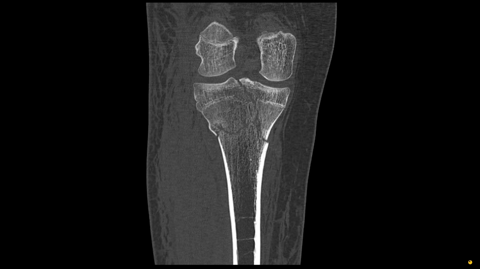
CT scan of the same patient showing the upper tibial comminuted fracture involving the articular surface.