
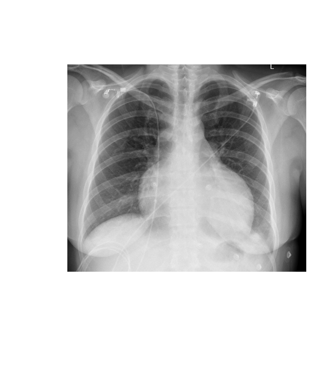
The following chest x-ray is from a 30 year old female patient who sustained a stab wound to her chest a few hours earlier. What do you see?
click to enlarge
The frontal chest x-ray shows an enlarged globular heart which indicates the presence of a pericardial effusion (bleeding into the pericardial sac in the context of the trauma). There are ECG dots and wires but otherwise, there is no visible foreign body. The lung fields appear clear.
Chest x-ray is not very sensitive in the diagnosis of pericardial effusion. A bedside ultrasound can aid in immediate diagnosis in the ED; otherwise, a formal ECHO should be obtained.
Some of the causes of pericardial effusion are: idiopathic, bacterial or viral infections, autoimmune disorders, uraemia, hypothyroidism, Dressler’s syndrome, malignancy and trauma. A globular heart in a dyspnoeic patient with any of the above risk factors should raise the suspicion of pericardial effusion.
[/peekaboo_content]