
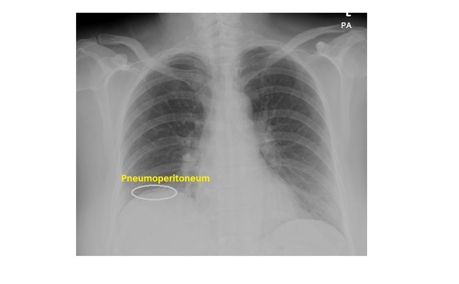
The following erect chest x-ray is from a 60 year old female who presented with a sudden onset of epigastric pain. The patient was also experiencing vague right-sided abdominal pain for a few days prior to the presentation. What can you note in the x-ray?

click to enlarge
The erect chest x-ray shows a subtle curvilinear lucency under the right hemi diaphragm, which is suggestive of free intraperitoneal air.
click to enlarge
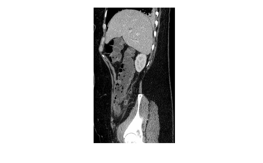
The following sagittal CT scan image shows free subdiaphragmatic air.
click to enlarge

click to enlarge
This patient had a retrocaecal appendicitis which perforated, causing pneumoperitoneum.
To allow the free air in the peritoneal cavity to rise, a patient needs to be upright for at least 10 minutes prior to obtaining the erect chest x-ray.
[/peekaboo_content]