
The following lateral soft tissue neck x-ray is from a 4 year old unwell child with a sore throat, fever and stiff neck. What can you deduce from the x-ray?

click to enlarge
The x-ray shows significant widening of the prevertebral soft tissue from C1 to C5, with up to 9 mm in front of the C2 vertebra. No air lucency/foreign body can be seen. The findings are highly suspicious for a retropharyngeal abscess.
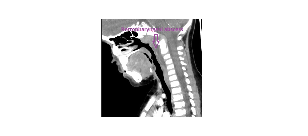
Post contrast sagittal and axial CT confirmed this diagnosis. The child was commenced on broad spectrum intravenous antibiotics and admitted under paediatric ENT services. I have no follow up on further management so I’m uncertain over whether the child underwent a surgical drainage.
click to enlarge

click to enlarge
CT scan with contrast is the preferred imaging method in a patient with suspected retropharyngeal abscess as it can differentiate between an abscess and cellulitis (to determine the need for surgical drainage). However, there is always a risk of radiation.
On a lateral neck x-ray, at the level of C2, the distance from the anterior surface of the vertebrae to the posterior border of the airway should be 7 mm or less, regardless of the patient’s age. At C6, this distance should be 14 mm or less in children younger than 15 years and less than 22 mm in an adult.
Reference: http://emedicine.medscape.com/article/995851
[/peekaboo_content]