
The following AP and lateral thoracolumbar junction x-rays are from a 38 year old man who has been involved in a MVA. What do you notice on the x-rays?

click to enlarge

click to enlarge
The thoracolumbar junction x-ray views show a compression fracture of L1 involving the superior vertebral end plate and the anterior body; there is also an undisplaced fracture of the spinous process of T12.
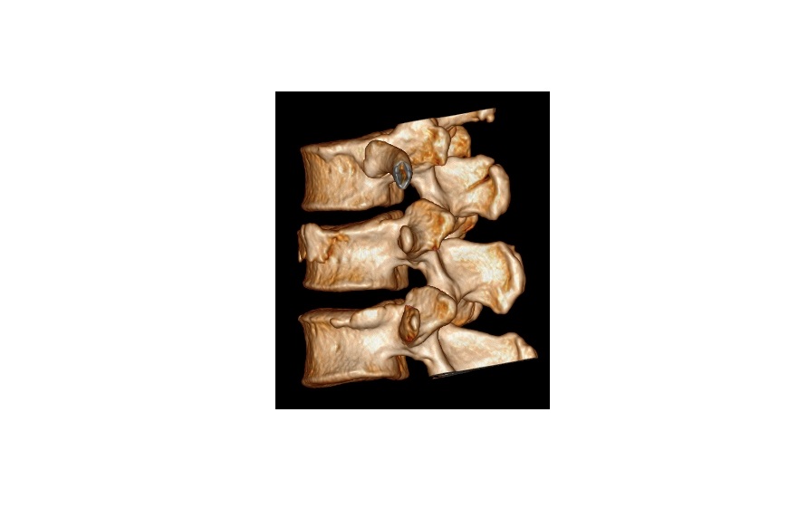
CT scans showed a compression fracture of L1 with no retropulsion of fragments. There was also a T12 spinous process fracture with an extension to the right facet joint.
This is a variant of a Chance fracture and is an unstable injury.
The patient did not have any abdominal/retroperitoneal injuries on CT and there was no neurological deficit. He was managed conservatively with a spinal brace.
click to enlarge
3D CT images from the same patient:

click to enlarge
click to enlarge
Chance fracture:
- Is a flexion-distraction injury related to lap seat belt use. It can also occur following a fall from a height.
- It is a horizontal fracture involving the anterior and posterior vertebral elements (vertebral body, pedicles and the spinous process)
- Tends to occur between T12 and L4.
- High incidence of gastrointestinal injuries (in up to 50% patients) involving the liver, spleen, duodenum or pancreas. Ecchymosis of the anterior abdominal wall should raise suspicion for the presence of this fracture.
- Be suspicious of compression fractures in young patients involved in an MVA.