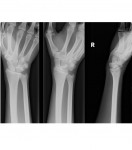
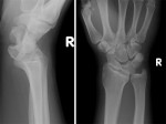
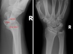
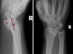
The following right wrist x-rays are from two different patients who each experienced a fall on their outstretched hand. Each show two different abnormalities so what can you deduce from the x-rays?
[peekaboo_link name=”Answer”]Answer[/peekaboo_link] [peekaboo_content name=”Answer”]
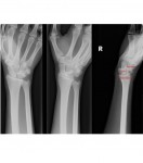
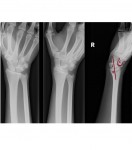
The first set of wrist x-rays show a perilunate dislocation whereas the second set exhibits a lunate dislocation.
Lunate and Perilunate dislocations:
- Common mechanism is a fall on the outstretched hand.
- Disruption of the ligament between the capitate and the lunate leads to dislocation of the capitate from the cup-shaped distal surface of the lunate.
- Normally in a lateral wrist view, the distal radius, the lunate and the capitate lie in a straight line. In a perilunate dislocation, the capitate and the metacarpals lie posterior to the line drawn through the distal radius and the lunate. However, in a lunate dislocation, the line drawn through the distal radius goes through the capitate but the lunate is volarly displaced.
- A lunate or perilunate dislocation can be diagnosed on an AP wrist view by noting a triangular lunate. Ordinarily, the lunate has a rhomboid shape on the AP view with parallel upper and lower borders.
- Risk with a lunate dislocation is acute carpal tunnel syndrome due to the compression of the median nerve by the volarly displaced lunate (incidence is approximately 25%)
- A perilunate dislocation is usually associated with other carpal bone fractures but most commonly with a transscaphoid fracture.
- Treatment for both types of dislocations is emergent closed reduction, splinting, followed by open reduction, ligament repair and internal fixation.
Normal lateral wrist alignment:
Perilunate dislocation:
Lunate dislocation:
My mnemonic is 3 P’s : Perilunate – Capitate – Posterior
References:
- Fundamentals of Diagnostic Radiology by Brant and Helms
- http://www.orthobullets.com/hand/6045/lunate-dislocation-perilunate-dissociation

