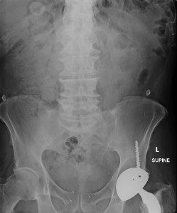
70 year old with vague upper abdominal pain with h/o previous ca prostate.He has diffuse tenderness in the Epigastrium and RUQ.
You obtain a plain x-ray abdomen as part of your initial investigations.
The supine abdominal film is attached.
What would you do next?
Supine abdominal film shows a nonspecific bowel gas pattern.There is no evidence of distended large/small bowel loops.There is faecal loading in the ascending colon.Patient has an artificial left hip joint and there are few ecg dots visble and ? implanted radiotherapy seeds in the pelvis.But if you look carefully,there is a rounded opacity with mild surrounding lucency in the RUQ.
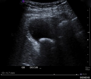
Based on anatomical location of the opacity,the likely cause is cholelithiasis.In an elderly patient with upper abdominal pain,this should raise the possibility of cholecystitis.How would you confirm this? Of course by requesting an ultrasound of the biliary tract.
Now,the ultrasound confirms your suspicion.There is a gall stone 20 mm in diameter in the neck of gall bladder.Patient had positive sonographic Murphy`s sign and the gall bladder wall is thickened,confirmimg the diagnosis of cholecystitis.
10-15 % of gall stones are visible on the xray due to calcium content.They have a variable position on abdominal xray depending on the gall bladder position.Often appear as rounded or clustered opacity in the RUQ.Their presence may be entirely incidental finding but should be part of differential diagnosis in a patient with abdominal pain.


