
A 70 year old male presents to ED complaining of shortness of breath. The patient has a history of pulmonary hypertension. An ECG is done:
Describe and Interpret the ECG
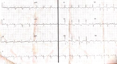
Rate: 102 b/min
Rhythm: Sinus tachycardia
Axis: Right Axis
Intervals
- PR 160ms
- QRS 120ms
- QT 430ms (Fridericia)
Additional:
P waves – tall P waves inferiorly >2.5mm meeting criteria for P pulmonale
QRS – RBBB Morphology, with fragmentation most noticeable in the inferior leads
T waves – inversion V1-V4 with ST depression
The above ECG shows a sinus tachycardia with a RBBB, P pulmonale and a strain pattern in the anterior leads. The fragmentation of the QRS is likely due to underlying scaring of the myocardium. These findings could be due to the patients underlying pulmonary hypertension, but other differential diagnosis include ACS, underlying right ventricular hypertrophy and acute PE.
In this clinical context further investigations to differentiate these causes include
- Obtaining an old ECG to decide if changes are old or new
- CXR to exclude pneumonia or other lung pathology causing the patients SOB/ strain pattern on the ECG
- D-dimer/Trop