
These elbow x-rays are from a 5 year old patient who experienced a fall onto her left elbow. What injuries can you note in the x-rays?
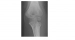
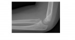
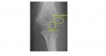
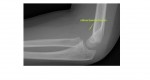
The elbow x-rays show a large effusion; there is an undisplaced lateral condylar fracture as well as a fracture through the capitellar ossification centre.
The patient was managed conservatively and did well.
Lateral condyle fractures are the second most common elbow injury in children (supracondylar fractures occur most frequently). These fractures can occasionally be confused with the developing ossification centres in children.
The usual order of appearance of ossification centres around the elbow can be memorised through the mnemonic CRITOL, for the first letter of each epiphysis. Knowing the age of appearance of these makes it possible to determine whether a bone fragment adjacent to the humerus represents a fracture or a normal ossification centre.
The approximate age of appearance of the centres are 2, 4, 6, 8, 10, and 12 years; this means that the lateral condylar ossification appears around 12 years of age. In this particular case, the age of the affected patient is only 5 years which implies that she has yet to develop an ossification for the lateral condyle. Thus, the bone fragment must represent a fracture.
Reference: Grainger and Allison’s Diagnostic Radiology- A Textbook of Medical Imaging
[/peekaboo_content]