
The AP wrist x-ray shows crowding of the carpal bones with a triangular configuration of the lunate (normally should look square).
 The lateral view shows dislocation of the lunate.
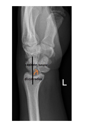
On lateral wrist x-ray, the distal radius, lunate and the capitate should form a straight line. In lunate dislocation, the distal radius and the capitate will remain in a straight line, but the lunate dislocates anteriorly.
Most important immediate complication is acute carpal tunnel syndrome due to compression of the median nerve in the carpal tunnel by the anteriorly dislocated lunate.
This patient underwent an initial successful closed reduction in ED, followed by internal fixation in the theatre.
Reference and further information on lunate dislocation can be found here