
The frontal chest x-ray shows a cavity in the left lung with adjacent area of consolidation.
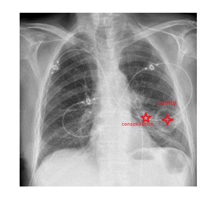
The patient had a CT scan of the chest which revealed a large cavitating mass in the left lower lobe. Biopsy revealed a squamous cell lung carcinoma.
Squamous cell lung cancer can produce parathyroid hormone related peptide which leads to hypercalcaemia.
Among different types of lung cancers, squamous cell carcinoma is the most likely cell type to show cavitation.
Reference: Grainger & Allison’s Diagnostic Radiology E-Book