
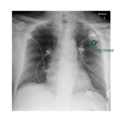
The chest x-ray shows a peripherally based lung mass in the left upper lobe. This pathology is nearly hidden under the ECG wires.
The patient, who had no significant past medical history, underwent a CT scan of their head for investigation of confusion. CT scan showed cerebral metastatic lesion with surrounding oedema.
Other causes of confusion to consider in a patient with lung carcinoma is hyponatraemia due to SIADH related to paraneoplastic syndrome (small cell carcinoma) & hypercalcaemia in squamous cell lung carcinoma.