
This left knee x-ray is from a 40 year old man with knee pain who experienced a twisting injury to the knee a few weeks ago. What can you see?

click to enlarge
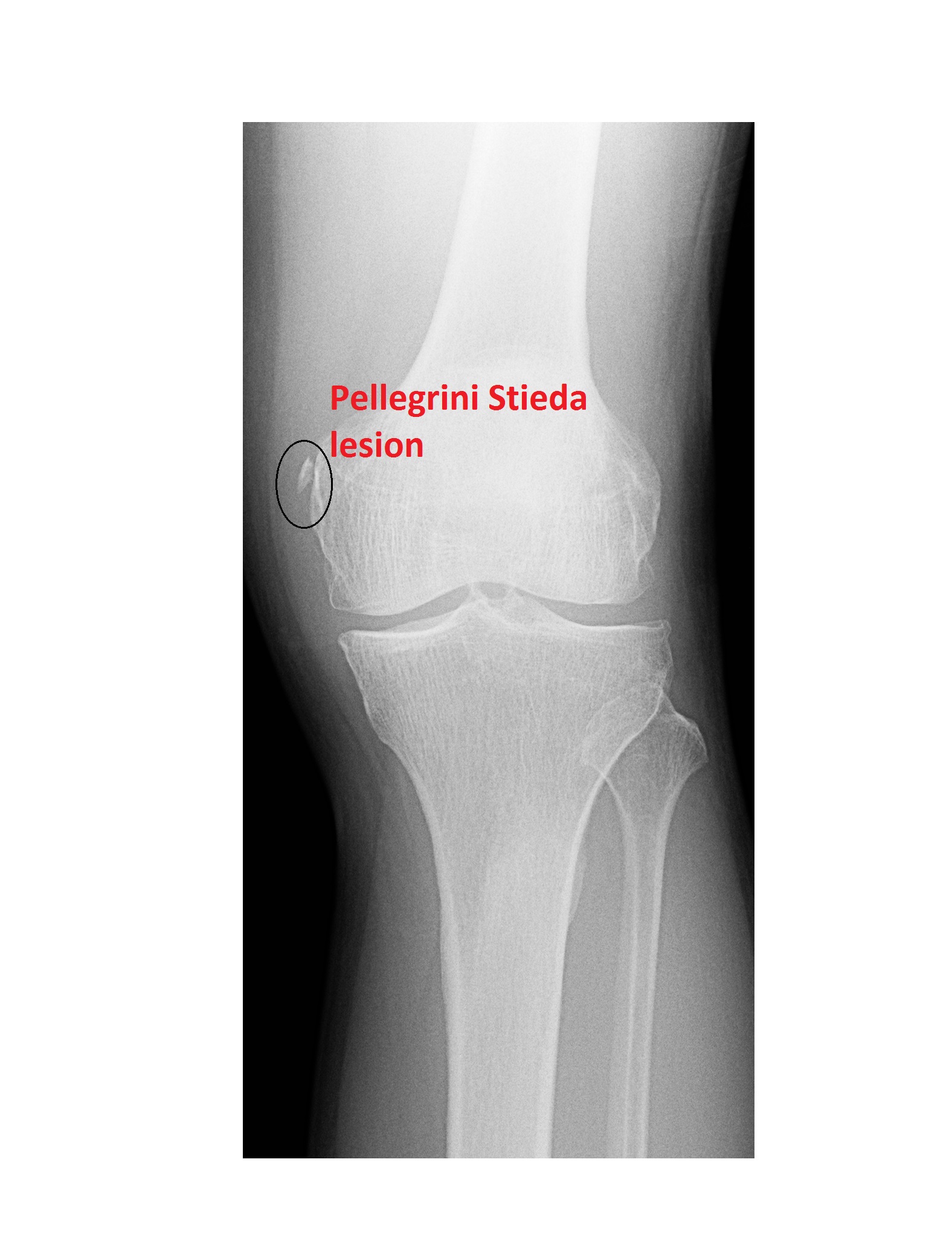
The knee x-ray shows an ossific density adjacent to the medial aspect of the medial femoral condyle; this is a Pellegrini Stieda lesion.
click to enlarge
Pellegrini Stieda lesion:
- An ossification adjacent to the MCL (medial collateral ligament) near the medial femoral condyle.
- The lesion is believed to be the result of past healed trauma such as Stieda’s fracture, which is an avulsion injury from the medial femoral condyle at the origin of the MCL.
- MR studies locate the ossifications deep within the superficial layer of the MCL.
- Treatment of isolated MCL tear is usually conservative.
reference: http://emedicine.medscape.com/article/89890
[/peekaboo_content]