Lab case 275 Interpretation

23 years old female presents to emergency department with c/o neck pain and upper thoracic cage pain for last 2 weeks. Patient has been taking regular analgesia with temporary relief. No significant past medical history. Otherwise well. No trauma.

HR 90, BP 117/80, afebrile, RR 18, sats 100 RA.

chest clear, abdomen soft non tender. Patient was discharged home after brief work up in ED and advised to follow up with GP. Continue reading

Lab case 276

23 years old female presents to emergency department with c/o neck pain and upper thoracic cage pain for last 2 weeks. Patient has been taking regular analgesia with temporary relief. No significant past medical history. Otherwise well. No trauma.

HR 90, BP 117/80, afebrile, RR 18, sats 100 RA.

chest clear, abdomen soft non tender. Patient was discharged home after brief work up in ED and advised to follow up with GP. Continue reading

Lab case 273 interpretation:

75 years old male, brought to ED by family with increasing lethargy and generalized weakness for last few days. 2 days ago patient had a fall in shower , seen by GP same day and was advised to observe.  No obvious injuries found.  His past med history includes T2DM, HTN, CKD. He is visiting from INDIA and has been intermittently non compliant with his medications.

GCS 13, HR 75, BP 85 systolic , temp 35.5. Peripherally shut down.

ECG : looks ischemic with STE AVR , and global STD. Following VBG is obtained: Continue reading

Lab case 273.

75 years old male, brought to ED by family with increasing lethargy and generalized weakness for last few days. 2 days ago patient had a fall in shower , seen by GP same day and was advised to observe.  No obvious injuries found.  His past med history includes T2DM, HTN, CKD. He is visiting from INDIA and has been intermittently non compliant with his medications.

GCS 13, HR 75, BP 85 systolic , temp 35.5. Peripherally shut down.

ECG : looks ischemic with STE AVR , and global STD. Following VBG is obtained: Continue reading

Ultrasound Post 3 Interpretation

30 year old male presents to ED with onset of right sided chest pain 2 hours ago. pain is pleuritic in nature and is associated with some Shortness of breath. His Vitals are HR 90, BP 130/70, RR 18, Sats 96 RA. He has history of primary spontaneous pneumothorax on right side about a year ago ,was treated with ICC insertion and subsequently resolved . Bedside ultrasound is performed and following images and clips are obtained.

Answer 1.  First Clip is of left side of chest ( normal side) . It shows 2 rib shadows, with pleural interface as bright white line. We can appreciate “ant crawling ” effect over pleural interface which is actually lung sliding and indicates absence of pneumothorax. Small vertical flashing lines intermittently are also evident which are called Comet tail artefacts.

Remember lung sliding can be absent in chronic lung conditions e.g severe emphysema, bronchiectasis, intubated patient.

Second clip is of symptomatic right side. It again shows rib shadows with bright line ( pleural interface ) , but it is quite static as compared to other side and we  can not appreciate ant crawling effect or lung sliding. Also there are no comet tail artefacts seen. This strongly indicates presence of pneumothorax on right side. There is also small movement coming towards centre like a curtain with breathing , it is called lung point.

Answer 2:

These images are  taken with M mode. First image shows sea shore sign , and second image shows bar code sign. Barcode sign indicates pneumothorax but is not very sensitive or specific .

Answer : 3. This clip shows ” lung point” which is highly sensitive for presence of pneumothorax. It appears as a curtain moving towards middle as the patient breaths and represents normal lung sliding coming in contact with abnormal lung.


Summary: 

Absence of lung sliding, and comet tail artefacts and presence of lung point strongly suggests presence of pneumothorax.

Ultrasound Post 3

30 year old male presents to ED with onset of right sided chest pain 2 hours ago. pain is pleuritic in nature and is associated with some Shortness of breath. His Vitals are HR 90, BP 130/70, RR 18, Sats 96 RA. He has history of primary spontaneous pneumothorax on right side about a year ago ,was treated with ICC insertion and subsequently resolved . Bedside ultrasound is performed and following images and clips are obtained. Continue reading

Ultrasound Post 2 Interpretation.

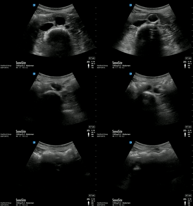
A registrar happens to do bedside ultrasound on a 55 years old male with abdominal pain and obtains following images.


Answers:

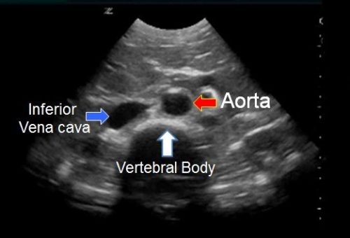
  1. Above is transvers view of distal aorta , and curvilinear probe has been used. Curvilinear probe is low frequency probe and gives higher depth so ideal for looking at deeper structures hence used in AAA/ AFAST scans.
  2. Hypoechoic ( black)  half circle at the bottom of image is vertebral body shadow which is the main landmark for identifying abdominal aorta. Remember ultrasound waves do not pass through the bones and you get a  black shadow as above . Other example is rib shadow when doing FAST scan.

The round structure on right of image ( left of patient ) is Aorta , while the elliptical structure on left of image ( right of patient ) is IVC. Following points will help further to differentiate between two.

a) Aorta lies on left of  while IVC lies on right of vertebral body ( patient ).

b) Aorta appears round in shape, while IVC appears elliptical mostly like a tear drop.

c) Both structures can be pulsatile so do not get trapped.

Below is coloured demonstration of above structures.